In brief: What is the structure of hair and how does it grow? InformedHealth org NCBI Bookshelf
Table Of Content

First, let’s see the microscopic image of an animal’s hair and try to identify its following features. All the following microscopic features are well identified in the hair-labeled diagram. Similar to the skin, hair gets its color from the pigment melanin, produced by melanocytes in the hair papilla. Different hair color results from differences in the type of melanin, which is genetically determined. As a person ages, the melanin production decreases, and hair tends to lose its color and becomes gray and/or white. The cuticle is your hair’s protective layer, composed of overlapping cells — like fish scales or roof tiles, but facing downwards.
What Is Human Hair Made Of?
In this chapter, the basic anatomy and the amazing and complicated biology of the hair follicle is reviewed. Enhanced knowledge on the normal dynamics of the hair provides understanding the basis of how the follicle behaves during a disease. However recent progress in our understanding of the biology and pathology of hair follicles should lead more effective therapies for hair disorders. Hair shaft moisture is actually in the medulla and is held in the central part of the hair shaft. The cuticle (the outer layer of the hair shaft), serves among other things to allow moisture to come in and out of the medulla. The cuticle protects the medulla from harmful environmental agents , sun, wind, pollution chemicals and other toxin.
Anatomy and Physiology of Hair
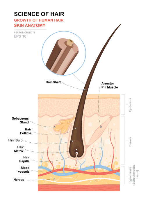
The hair shaft is the part of the hair not anchored to the follicle, and much of this is exposed at the skin’s surface. The rest of the hair, which is anchored in the follicle, lies below the surface of the skin and is referred to as the hair root. The hair root ends deep in the dermis at the hair bulb, and includes a layer of mitotically active basal cells called the hair matrix. The hair bulb surrounds the hair papilla, which is made of connective tissue and contains blood capillaries and nerve endings from the dermis (Figure 1). In the second stage of development, hair germ elongates into a cord of epithelial cells and forms the hair peg (stages 3 and 4). It is surrounded by mesenchymal cells that eventually transformed to the fibrous sheath.
Anatomy at a Glance
Figure 1 Fine long hair was detected all over the body. - ResearchGate
Figure 1 Fine long hair was detected all over the body..
Posted: Thu, 08 Feb 2018 16:50:31 GMT [source]
The middle layer of the hair shaft is called the cortex, made of keratin fibers. The strength, color and texture of a hair fiber are provided by the cortex layer of the hair shaft. This thin and colorless layer made up of between six to ten overlapping layers of long cell remnants, serves as a protection to the cortex.

But, the length and texture of the hair are different in the different regions of the animal or human body. This specialized immune environment of IP is required to prevent destructive immune reactions in critical regions. Other immune privileged sites include the anterior chamber of the eye, testis, brain and placenta. Hair follicle IP has a unique characteristic of recurring in a cyclic pattern. The hair follicle is a skin appendage located deep in the dermis of the skin. From the inside out, these are the medulla, cortex, and cuticle.
A network of blood vessels presents at the hair papilla supplies nutrients to the hair and helps them grow. This dermal papilla provides nutrition (blood supply) to the hair of an animal. A basement membrane separates the dermal papilla from the hair matrix. I hope you got the basic idea of the different microscopic features of the hair of animals. Now, you may learn the details of microscopic features of every single structure of the animal’s hair.
Bold Hold Collection
Hair follicles may alter the structure of the hairs they produce in response to circulating hormones. The cuticle is made from 6to 11 layers of overlapping semi-transparent keratin scales (which make thehair waterproof and allow it to be stretched). The cortex is made from tiny fibers of keratin running parallel to each other along the length of the hair shaft (as shown in the photo of the split hair end above). But as they move up through thefollicle, they begin to change shape, and a protein called keratin develops inside the cells.
The pigments that give your hair its natural color are tucked among these protein strands and protected from the elements by the translucent layer of cuticle cells. A healthy cuticle is more than just a protective layer – much of the shine that makes healthy hair so attractive is due to the cuticle. This, together with the pigment in the cortex (see below), gives hair its characteristic appearance. A normal cuticle has a smooth surface that allows light to penetrate to the cortex. But this smooth surface is vulnerable to damage from weather and chemical treatments.
Basic Hair Structure: Hair Follicle and Hair Shaft Function Explained
Eating nutritious foods is one way to improve hair from the inside out. Check with your doctor if you have questions on your hair’s growth and how it might be impacted by your health. Some people have curly corkscrew hair, while others have thick, straight, shiny hair. The medulla may be absent or continuous, amorphous or vacuolated with a narrow or broad width.
Central banks should resolve to adopt a more humble approach to central banking Mint - Mint
Central banks should resolve to adopt a more humble approach to central banking Mint.
Posted: Tue, 12 Dec 2023 08:00:00 GMT [source]
In the great majority of cases, however, it is simply a matter of aging. The hair matrix is the epithelial layer involved in hair production. When the superficial basal cells divide, they produce daughter cells that are pushed toward the surface as part of the developing hair. The medulla contains relatively soft and flexible soft keratin. Matrix cells closer to the edge of the developing hair form the relatively hard cortex. The cortex contains hard keratin, which gives hair its stiffness.
Hair growth begins with the production of keratinocytes by the basal cells of the hair bulb. As new cells are deposited at the hair bulb, the hair shaft is pushed through the follicle toward the surface. Keratinization is completed as the cells are pushed to the skin surface to form the shaft of hair that is externally visible. The external hair is completely dead and composed entirely of keratin. Furthermore, you can cut your hair or shave without damaging the hair structure because the cut is superficial. Most chemical hair removers also act superficially; however, electrolysis and yanking both attempt to destroy the hair bulb so hair cannot grow.
When the hair has separated completely from the papilla, the supply of blood is cut off in the final resting phase, which is also called the telogen phase. The hair is gradually pushed out of the skin and eventually falls out. The full strand of hair develops from this group of hardened hair cells. Because new hardened cells keep on attaching to the hair from below, it is gradually pushed up out of the skin. In this way, a single hair on your head grows at a rate of about 1 cm per month. Facial hair, and especially eyelashes, eyebrows and body hair grows at a slower pace.
The human hair is formed by divisions of cells at the base of the follicle. As the cells are pushed upward from the follicle’s base, they become keratinized (hardened) and undergo pigmentation. In addition, the hair labeled diagram shows a highly vascularized connective tissue layer that surrounds the outer root layer of the hair follicle. The microscopic figure of the hair follicle shows the basal lamina that separates the outer root layer from the connective tissue layer. This basal lamina is the glassy membrane of the hair follicle structure. The glassy membrane of the hair follicle structure is strongly eosinophilic.
This diagram shows the hair root that contains the cuticle, cortex, and medulla. It also shows the Huxley’s layer and Henle’s layer that form the inner root layer of the hair follicle. Again, the arrector pili muscle lies on the side of the hair follicle and forms an angle with the skin surface.
You now know all about the various structures which form the hair follicle and hair shaft. On the next page, learn how all these different parts of the hair follicle interact to create the hair on your head over and over again throughout life. Vitamin C is a powerful antioxidant and protects hair follicles from damaging free radicals that contribute to discoloration and even hair loss. Hair grows as your blood vessels deliver nutrients to the cells inside of the hair bulb. As hair cells grow, they are attached to the follicle and the older cells are pushed outwards resulting in the strand-like appearance of your hair.
Microscopically, you will find minute air bubbles both in the medulla and the cortex of the hair shaft. Each hair of an animal consists of two parts – one visible on the body surface and another part anchored in the thickness of the skin. The visible part of the hair is known as the shaft, and the embedded part is the hair root.


Comments
Post a Comment